 |
| SPLEEN |
INTRODUCTION
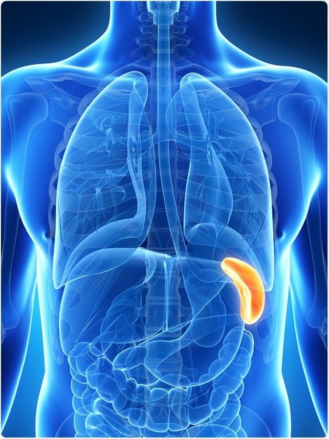
Spleen (Greek splen and Latin Lien) is a lymphatic organ connected to the blood vascular system. It acts as a filter for blood and plays an important role in the immune responses of the body.
Location


Location of spleen (a) in relation to the nine regions of abdomen (b) in relation to the fundus of stomach and the diaphragm
The spleen (Latin low spirits) is a wedge-shaped organ lying mainly in the left hypochondrium, and partly in the epigastrium. It is wedged in between the fundus of the stomach and the diaphragm. The spleen is tetrahedral in shape.
Dimensions
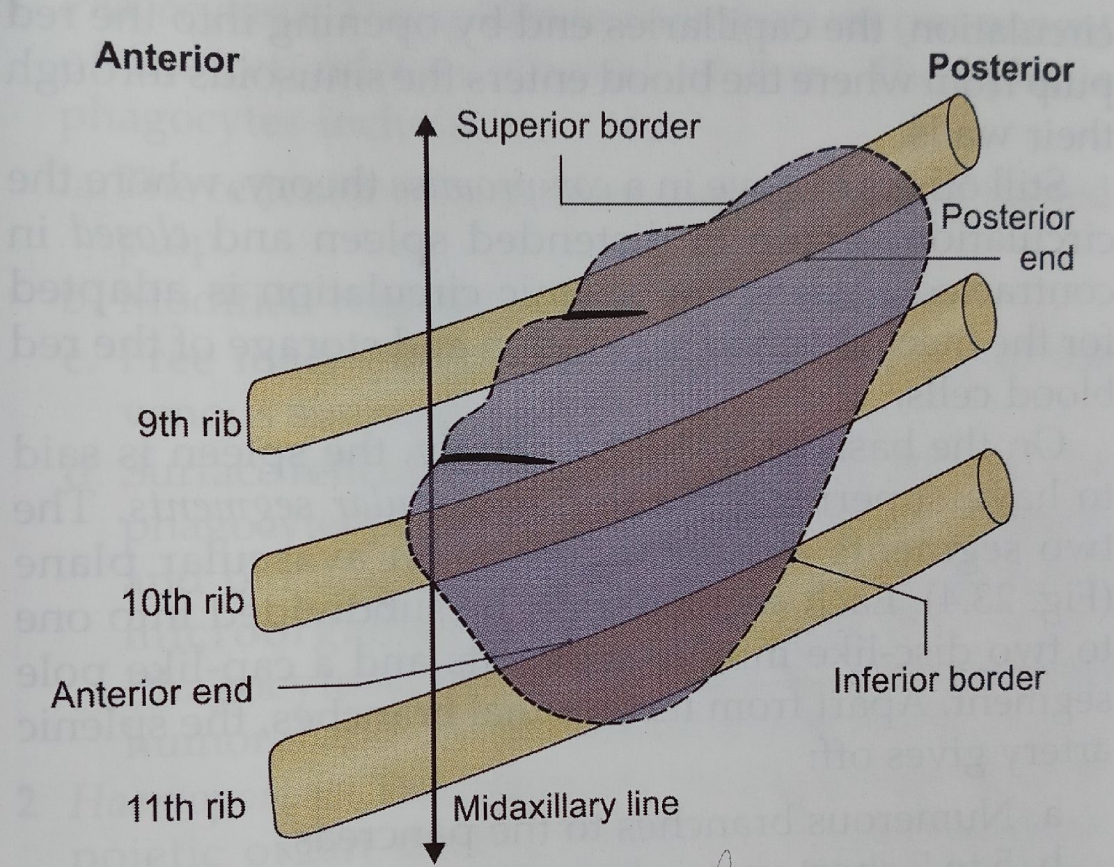
The spleen is soft, highly vascular and dark purple in colour. The size and weight of the spleen are markedly variable. On an average the spleen is 1 inch or 2.5 cm thick, 3 inches or 7.5 cm broad, 5 inches or 12.5 cm long,7 ounces in weight, and is related to 9th to 11th rihe The odd numbers are 1, 3, 5 are 7, 9, 11. Normally, the spleen is not palpable.
Position (Axis of Spleen)
 |
| Position of spleen in relation to left 9th - 11th ribs |
The spleen lies obliquely along the long axis of the 10th rib. Thus it is directed downwards, forwards and laterally, making an angle of about 45 degrees with the horizontal plane.
External Features
The spleen has two ends, three borders, two surfaces, two angles and hilum.
Two Ends
• The anterior or lateral end is expanded and is more like a border. It is directed downwards and forwards, and reaches the midaxillary line.
• The posterior or medial end is rounded. It is directed upwards, backwards and medially, and rests on the upper pole of the left kidney.
Three Borders
• The superior border is characteristically notched near the anterior end.
• The inferior border is rounded.
• The intermediate border is also rounded and is directed to the right.
Two Surfaces
• The diaphragmatic surface is convex and smooth.
• The visceral surface is concave and irregular.
Two Angles
Anterobasal angle: It is the junction of superior border with lateral or anterior end. It is the most forwand border Anterobasal angle: It is the junction of superior projecting part of spleen. When spleen is enlarged, this is felt first, so this is called as "clinical angle of spleen. Posterobasal angle: Junction of inferior border with lateral or anterior end of spleen.
Hillum
Hilum lies between superior and intermediate borders. It is pierced by branches and tributaries of splenic vessels.
Relations
Peritoneal Relations
 |
| Peritoneal ligaments attached to the spleen and common sites of accessory spleen |
The spleen is surrounded by peritoneum, and is suspended by following ligaments.
1 The gastrosplenic ligament extends from the hilum of the spleen to the greater curvature of the stomach. It contains the short gastric vessels and associated lymphatics and sympathetic nerves.
2 The lienorenal ligament extends from the hilum of the spleen to the anterior surface of the left kidney. It contains the tail of the pancreas, the splenic vessels, and associated pancreaticosplenic lymph nodes, lymphatics and sympathetic nerves.
3 The phrenicocolic ligament is not attached to the spleen, but supports its anterior end. It is a horizontal fold of peritoneum extending from the splenic flexure of colon to the diaphragm, opposite the 11th rib in the midaxillary line. It limits the upper end of the left paracolic gutter.
Visceral Relalions
 |
| Visceral relations of spleen |
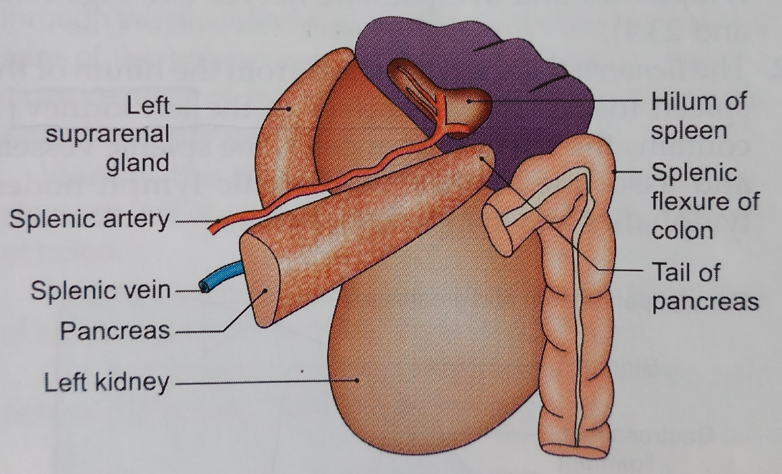
Visceral surface
The visceral surface is related to the fundus of the stomach, the anterior surface of the left kidney, the splenic flexure of the colon and the tail of the pancreas. The gastric impression, for the fundus of the stomach, lies between the superior and intermediate borders. It is the largest and most concave impression on the spleen. The renal impression, for the left kidney, lies between the inferior and intermediate borders. The colic impressions, for the splenic flexure of the colon, occupies a triangular area adjoining the anterior end of the spleen. Its lower part is related to the phrenicocolic ligament. The pancreatic impression, for the tail of the pancreas, lies between the hilum and the colic impression. The hilum lies on the inferomedial part of the gastric impression along the long axis of the spleen. It transmits the splenic vessels and nerves, and provides attachment to the gastrosplenic and lienorenal ligaments.
Diaphragmatic Surface
The diaphragmatic surface is related to the diaphragm which separates the spleen from the costodiaphrag- matic recess of pleura, lung and 9th, 10th and 11th ribs of the left side.
Blood Supply Of Spleen
 |
| Blood supply of spleen |
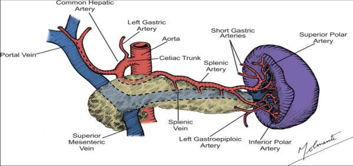
Arterlal Supply
The spleen is supplied by the splenic artery which is the largest branch of the coeltac trunk. The artery is tortuous in its course to allow for movements of the spleen. It passes through the lienorenal ligament to reach the hilum of the spleen where it divides into fu or more branches. These branches enter the spleen supply it. Within the spleen it divides repeatedly to form successfully the straight vessels called penicilli, which further divide into ellipsoids and arterial capillaries Further course of the blood is controversial. According to closed theory of splenic circulation, the capillaries are continuous with the venous sinusoids that lie in the red pulp; the sinusoids join together to form veins. However, according to open theory of splenic circulation, the capillaries end by opening into the red pulp from where the blood enters the sinusoids through their walls. Still others believe in a compromise theory, where the circulation is open in distended spleen and closed in contracted spleen. The splenic circulation is adapted for the mechanism of separation and storage of the red blood cells. On the basis of its blood supply, the spleen is said to have superior and inferior vascular segments. The two segments are separated by an avascular plane. Each segment may be subdivided into one to two disc-like middle segments and a cap-like pole segment. Apart from its terminal branches, the splenic artery gives off:
a. Numerous branches to the pancreas,
b. 5 to 7 short gastric branches, and
c. The left gastroepiploic artery.
Venous Drainage
The splenic vein is formed at the hilum of the spleen. It runs a straight course behind the pancreas It joins the superior mesenteric vein behind the neck of the nancreas to form the portaf vein. Its Iibutaries are the hort gastric. Teft gastroepiploic, pancreatic and inferior mesenteric veins.
Lymphatic Drainage
Splenic tissue proper has no lymphatics. A few Iymphatics arise from the connective tissue of the capsule including trabeculae and drain into the pancreaticosplenic lymph nodes situated along the splenic artery.
Nerve Supply
Sympathetic fibres are-derived from the coeliac plexus. They are vasomotor in nature. They atso supply some smooth muscle present in the capsule.
Functions of the Spleen
1 Phagocytosis: The spleen is an important component of the reticuloendothelial system. The splenic phagocytes include:
a. The reticular cells and free macrophages of the red pulp.
b. Modified reticular cells of the ellipsoids.
c. Free macrophages and endothelial cells of the venous sinusoids, and
d Surface reticular cells of the lymphatic follicle. The phagocytes present in the organ remove cell debris and old and effete RBCS, other blood cells and microorganisms, and thus filter the blood. Phagocytosis of circulating antigens initiates humoral and cellular immune responses
2 Haemopoiesis: The spleen is an important haemo- poietic organ during foetal life. Lymphopoiesis continues throughout life. The lymphocytes manufactured in it take part in immune responses of the body In the adult spleen, haemopoiesis can restart in ertain diseases, like chronic myeloid leukaemia and myelosclerosis.
3 Immune responses: Under antigenic stimulation, there occurs increased lymphopoiesis for cellular responses, and increased formation of plasma cells for the humoral responses.
4 Storage of RBCS: Red blood cells can be stored in the spleen and released into the circulation when needed. This function is better marked in animals than in man.
HISTOLOGY
 |
| Histology of spleen |
Histologically, spleen is made up of the following four component parts.
1 Supporting fibroelastic tissue, forming the capsule, coarse trabeculae and a fine reticulum. In human, the smooth muscle cells in the capsule and trabeculae are few, and the contraction and distension of spleen are attributed to constriction or relaxation of the blood vessels, which regulate the blood flow in the organ.
2 White pulp consisting of lymphatic nodules arranged around an arteriole called malpighian corpuscle.
3 Red pulp is formed by the collection of cells in the interstices of reticulum, in between the sinusoids. The cell population includes:
a. All types of lymphocytes (small, medium and large)
b. All three types of blood cells (RBC, WBC and platelets)
c. The fixed and free macrophages. Lymphocytes are freely transformed into plasma cells which can produce large amounts of antibodies the immunoglobulins.
4 Vascular system transverses the organ and permeates it thoroughly.
Source BD CHAURASIA'S Anatomy
Comments
Post a Comment